How Is Implant Treatment Planned for a Single Missing Tooth?
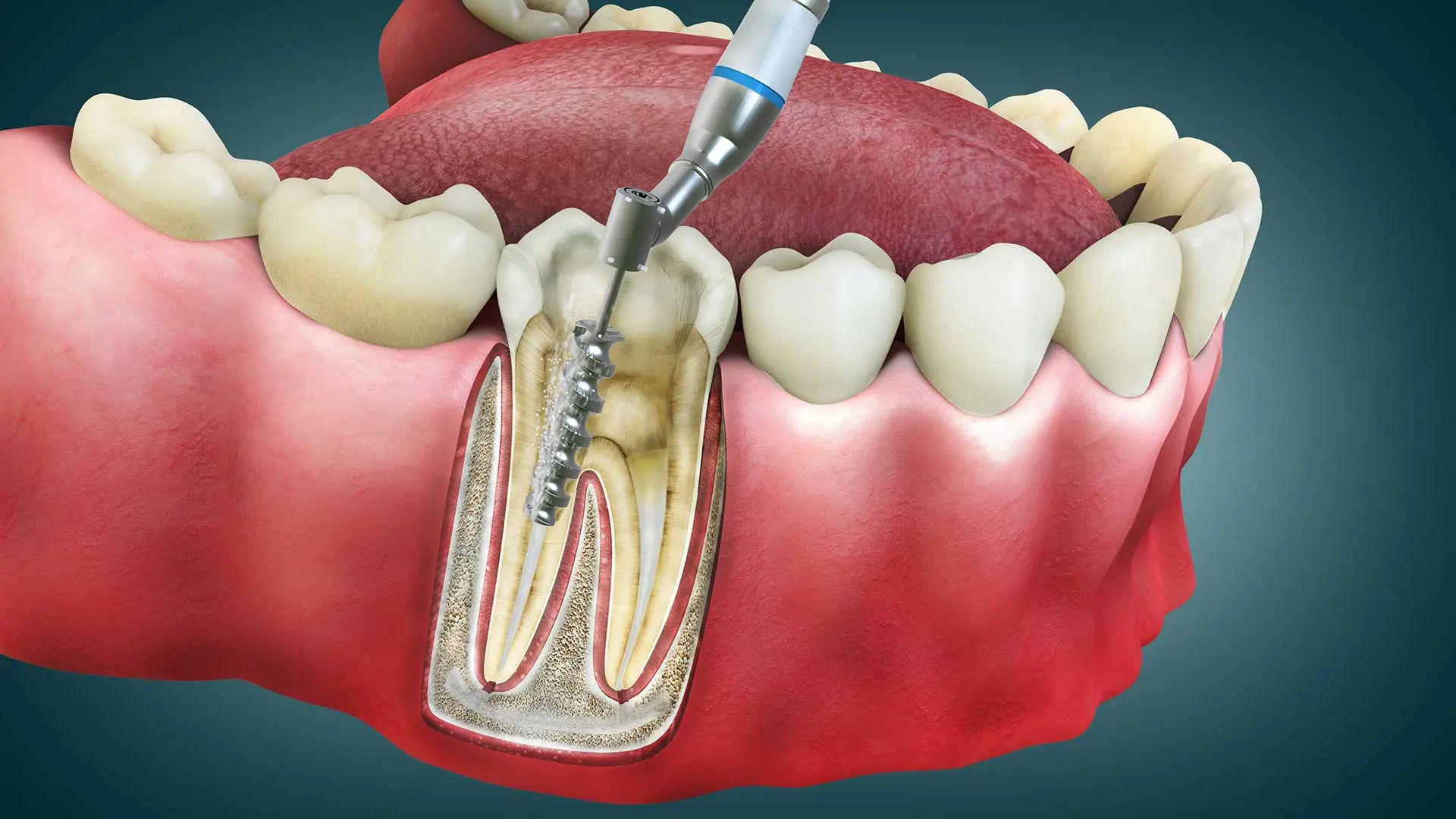
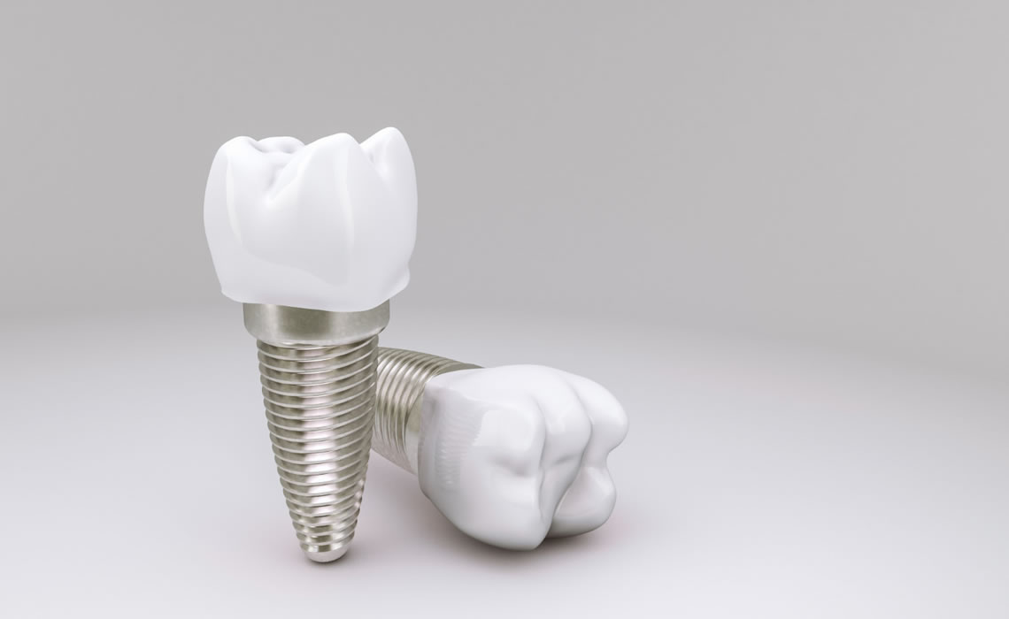
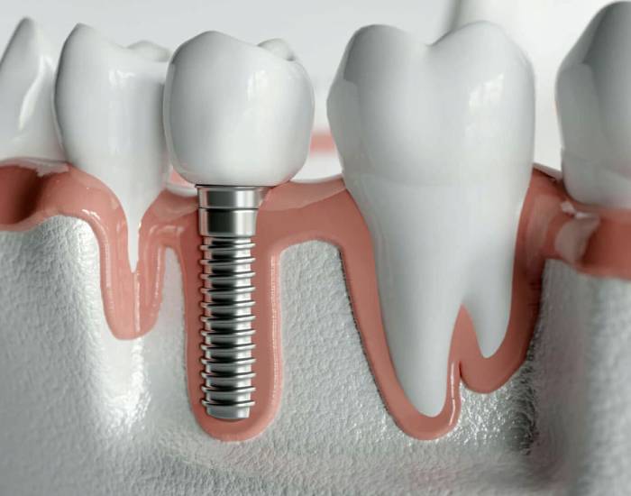
When planning implant treatment for a single missing tooth, the individual’s intraoral anatomy is examined using three-dimensional imaging technologies, and the bone density in the area where the titanium root will be placed is calculated. In light of this data, the physician digitally designs the angle, diameter, and length of the artificial root to be placed in the gap, without intervening on the other teeth.
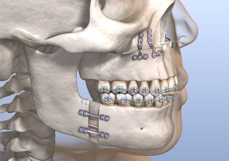
In dental practice, treatment planning is the fundamental stage for ensuring the long-term functional and anatomical sustainability of the outcome to be achieved. Jaw surgery specialists map the structure of the jawbone down to the millimeter as a result of radiological evaluations. Thanks to this mapping, the exact position in which the material that will replace the missing tooth will sit is determined before the surgical intervention. The fact that the application covers only a single tooth is based on the principle of preserving the neighboring dental tissues.
The main considerations taken into account during the planning stage can be summarized as follows:
- Taking the patient’s general state of health and the medical history of any medications they use,
- Assessing the current health status of the gums through a clinical examination,
- Measuring the thickness and height of the bone via three-dimensional radiographic scans,
- Analyzing the distribution of chewing forces across the jaw,
- Calculating the distance from the artificial root to be placed to the surrounding nerve canals and anatomical cavities.
What Is Done During the Pre-Treatment Imaging and Measurement Process?
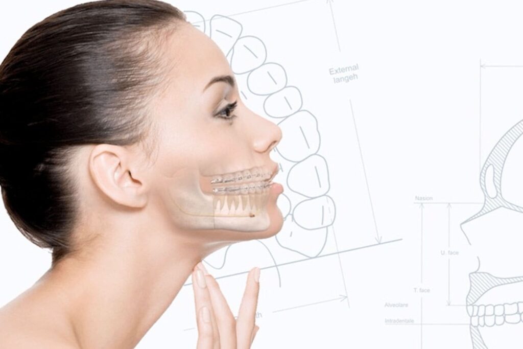
During the pre-treatment imaging and measurement process, the width and height of the jawbone and its distance to the nerve canals are measured down to the millimeter using panoramic X-rays and dental tomography (CBCT). The three-dimensional data obtained allows the position of the artificial root to be placed to be determined in advance in a way that will not damage the surrounding tissues.
The advancement of imaging technologies has significantly increased treatment predictability in dental practice. Two-dimensional X-rays alone can sometimes fail to provide sufficient data on the thickness of the jawbone. Three-dimensional dental tomography, which comes into play at this point, allows the bone tissue to be examined in cross-sections. By examining these cross-sections on a computer, the physician selects the diameter and length of the titanium screw in a form compatible with the individual’s own anatomy.
| Imaging Method | Purpose of Use | Data Obtained |
|---|---|---|
| Panoramic X-ray | General examination of the entire jaw structure, teeth, and joints in a single plane. | The general condition of the tooth roots, the existing bone level, and a two-dimensional view of the jaw anatomy. |
| Dental Tomography (CBCT) | Detailed volumetric analysis of the targeted area and identification of the nerve pathways. | The three-dimensional volume and density of the bone tissue, and the exact millimeter distance to the nerve canals. |
| Digital Intraoral Scanning | Taking measurements of the teeth with optical cameras for the prosthesis (crown) stage. | A three-dimensional model of the intraoral soft tissue and neighboring teeth, transferred to a computer environment. |
How Does Single Tooth Implant Planning Work at Istanbul OPC Clinic?
Within OPC Clinic, which serves the Istanbul area, single tooth implant planning proceeds by starting with a comprehensive intraoral examination and continuing with the transfer of the patient’s anatomical data to digital systems, thereby creating an individualized treatment protocol. Every step is carried out with standardized medical procedures, taking the individual’s physiological structure into account.
For individuals applying to the clinic, the first step is a preliminary examination in which the general condition of the intraoral tissues is assessed. After listening to the complaints of patients in Istanbul, OPC Clinic physicians request the necessary radiographic examinations. After the tomography data is reviewed, it is determined whether there is sufficient bone infrastructure for placing the artificial root. The process continues with patient consent and information. On the day of the application, under sterile conditions, medical titanium is placed in the targeted area using only local anesthesia, and the wound site is closed, moving on to the healing process.
Planning covers not only the surgical portion but also the prosthesis stage that will follow, from the very first day. In line with digital records taken with intraoral scanners, once the artificial root has fused with the bone, the color, size, and shape of the tooth that will go on top of it are designed from the outset to be compatible with the individual’s other teeth. This holistic approach ensures that function and aesthetics are carried out together.
How Does Planning Change in the Case of Insufficient Jawbone?
In the case of insufficient jawbone, the planning stage includes additional surgical procedures involving the addition of biologically compatible bone powder (graft) to the area before the artificial root is placed. This additional procedure adds a new healing period to the treatment schedule in order to create sufficient tissue volume for the titanium structure to be supported.
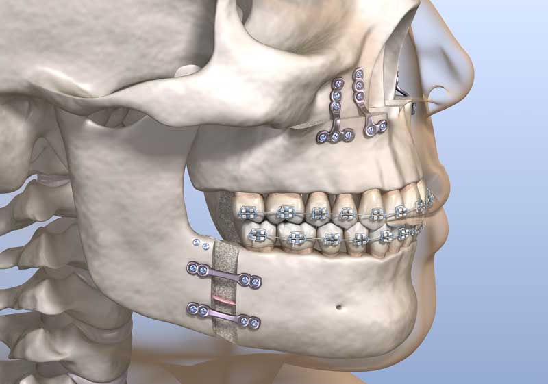
When a tooth is extracted, the bone tissue in that area loses the stimulating effect coming from chewing forces. Over time, this lack of stimulation results in bone resorption and a reduction in volume. Bone deficiency is a situation frequently encountered by physicians, especially when planning for individuals who lost their tooth many years ago. At this point, jaw surgery turns to the structural procedures we call bone grafting.
- Bone Powder (Graft) Application: The placement of synthetic or animal-derived sterile bone minerals in the area where volume has decreased.
- Use of a Membrane: Covering the area with a special barrier in order to prevent the added bone powder from dispersing and to ensure that bone cells, rather than gum cells, proliferate in that area.
- Sinus Floor Elevation: In the case of sagging of the anatomical air cavities (sinuses) located in the rear areas of the upper jaw, this is the procedure of carefully pushing the sinus membrane upward so that bone powder can be added.
According to Which Criteria Is Implant-Supported Prosthesis Planning Carried Out?
Implant-supported prosthesis planning is the process of designing, in a laboratory setting, the crown to be produced from porcelain or zirconium materials, based on anatomical criteria such as the color tone and shape of the neighboring teeth and the relationship between the upper and lower jaw bite. This design takes its final form once the intraoral measurements are taken after the fusion of the artificial root with the bone is complete.
No matter how precisely the surgical stage is carried out, the part the individual sees and uses in daily life is the prosthesis — that is, the crown. For this reason, the planning of the prosthesis stage involves just as much detail as the surgery. Models prepared using digital measurement techniques are transferred to devices that mimic jaw movements (articulators). In this way, the pressure that the newly made artificial tooth will exert on the opposing tooth during chewing is calculated. Since excessive pressure or insufficient contact can lead to jaw joint problems, millimeter-level adjustments are carried out in the laboratory setting.
| Material Used | Physical Properties | Area of Use |
|---|---|---|
| Zirconium-Based Porcelain | A white metal substructure with light transmission close to that of a natural tooth, high tissue compatibility, and an aesthetic appearance. | Generally missing teeth in the front area, where aesthetic expectations are high. |
| Metal-Based Porcelain | Porcelain applied over a classic gray metal substructure, quite resistant to chewing forces. | Missing teeth in the rear area (molars) where the chewing load is intense. |
| Monolithic Zirconium | A high-strength structure produced as a single block, without an extra layer of porcelain on top. | Specific cases where aesthetics and extra durability are expected at the same time. |
How Is the Healing Process Reflected in the Treatment Schedule?
The healing process is reflected in the treatment schedule as part of the average two-to-six-month waiting period needed for the titanium material to integrate with the jawbone cells (osseointegration). Throughout this period, the physician monitors the course of tissue fusion by performing radiographic check-ups at certain intervals.
Immediately after the surgical procedure, bone cells (osteoblasts) begin to proliferate by attaching to the titanium surface. This biological event forms the basis of the treatment applied and requires a certain amount of time. After the operation, the physician provides the patient with a roadmap for the healing period. This roadmap includes details such as suture removal in the first week, the use of antibiotics and painkillers, and how to maintain oral hygiene. In cases where bone powder is used, this waiting period is entered into the schedule as extending a few months longer than a standard procedure.
Which Specialties Work Together in Single Tooth Implant Planning?
In single tooth implant planning, oral, dental, and maxillofacial surgery specialists generally work together with prosthetic dentistry specialists, adopting a multidisciplinary approach. While the surgical department is responsible for placing the artificial root into the jawbone at the appropriate angle, the prosthesis specialist ensures the functional and aesthetic compatibility of the crown that will be placed on top.
The secret to a successful application in dentistry lies in bringing together the accumulated knowledge of different specialty areas. Just as it is important to place the root in the correct location, it is equally important for the shape of the tooth to be made on top of it to be compatible with the jaw’s bite. In well-equipped centers such as OPC Clinic in Istanbul, the patient’s radiographic data is jointly evaluated by both disciplines. When necessary, gum (periodontology) specialists are also included in the planning, ensuring the health of the gum tissue surrounding the artificial root is guaranteed.
Frequently Asked Questions
1. Is there an age limit for the treatment of a single missing tooth?
For the treatment to be applicable, the individual is expected to have completed the development of their jawbone. This generally corresponds to ages 16-17 in women and 18-19 in men. No upper age limit is specified, as long as the general state of health remains suitable.
2. How is the type of anesthesia determined during the planning process?
The great majority of procedures are planned to be carried out using the local anesthesia method, in which only the relevant area of the patient is numbed. However, in patients with an advanced dental phobia or gag reflex, general anesthesia or sedation alternatives are considered.
3. Why is gum health important in the area where the implant will be placed?
The gums serve as a biological barrier that protects the artificial root against infections. In areas where gum disease is present, gum treatment is planned first, and surgical planning proceeds once the tissues have reached a healthy form.
4. Is the fabrication of a temporary tooth included in the planning?
Especially in gaps that occur in the front group of teeth, temporary crowns prepared to not be exposed to chewing force during the healing process can be included in the planning in order to address aesthetic concerns.
5. How is treatment planning carried out for diabetic patients?
In diabetic patients, blood values such as HbA1c are requested to be measured before treatment. In individuals whose blood sugar levels are under control and within the reference ranges determined by the physician, planning proceeds in accordance with standard procedures.
6. How is bone development radiologically confirmed?
In individuals of growing age, hand-wrist radiographs are requested to examine the state of the growth plates, in order to understand whether bone development has been completed. If the plates have closed, the surgical stage can proceed.
7. How much time does the operation take up in the treatment schedule?
The surgical operation, which covers the placement of a single artificial root, is scheduled to be completed in a clinical setting within an average of 30 to 60 minutes, including the duration of the local anesthesia.
8. How is the process managed for individuals taking blood thinners?
Patients taking blood thinner medication are assessed as a result of a joint consultation between the dentist carrying out the treatment and the medical doctor who prescribed the medication. When necessary, planning is carried out by adjusting the medication dosage a few days before the procedure.
9. Does the titanium material used cause allergies?
Titanium is a material with an extremely high level of compatibility with human biology. Just as it is used in many branches of medicine, including orthopedics, its use in dentistry is also quite widespread, and the likelihood of it being perceived as a foreign substance by the body is very low.
10. Does smoking affect the planning process?
Smoking can slow down wound healing by negatively affecting blood circulation in the intraoral tissues. Physicians ask their patients to stay away from tobacco products for a certain period covering before and after the treatment.
11. How long does the laboratory stage take in prosthesis planning?
Once bone fusion is complete, the intraoral measurements are sent to the laboratory. The individualized production of zirconium or porcelain crowns, the fittings, and reaching the final form are completed within an average of 7 to 14 days, depending on clinical conditions.
12. How should the eating routine be planned after the procedure?
In order to protect the sutures and soft tissues at the operation site, eating warm and soft-textured foods is planned for the first few days. Hard, crusty, and overly hot foods are removed from the diet throughout the healing process.
13. At what intervals are check-up appointments scheduled?
One week after the surgical stage, the first appointment is given for suture removal and a check-up. Afterward, radiographic check-ups are carried out at the 1st and 3rd months in order to monitor the bone-formation process known as osseointegration.
14. What is the difference between planning an implant versus a bridge prosthesis?
With bridge prostheses, the healthy neighboring teeth need to be reduced in size in order to form a body in place of the missing tooth. In the implant procedure, on the other hand, no mechanical procedure is applied to the neighboring teeth at all, and only the area with the gap is treated.
15. Is a panoramic X-ray alone sufficient for planning?
In many cases, a panoramic X-ray provides sufficient data in terms of showing the general condition, while in specific situations where the anatomical structures are complex or where the bone density requires detailed measurement, three-dimensional tomography is needed.