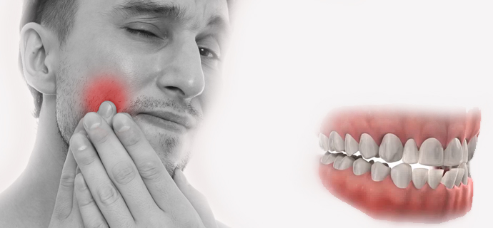
You’ve noticed your jaw aches, perhaps you hear clicking sounds, or you can’t open your mouth as easily as before, and you’ve decided to see a doctor to solve the problem. But which doctor should you see for jaw pain? Orthopedics, ENT, neurology?
The correct answer is to see a temporomandibular joint (TMJ) specialist. Doctors specializing in this area may come from different medical or dental disciplines and manage different aspects of treatment. However, the doctor who will diagnose jaw pain and dysfunction is either an oral and maxillofacial surgeon or a prosthetic dentistry specialist (prosthodontist) experienced in TMJ issues. Ideally, both specialists should examine you together.
How is a Temporomandibular Joint Examination Performed Step-by-Step?
During the initial examination, we ask the patient detailed questions: the onset of symptoms, the severity of pain, and the extent of jaw function loss are assessed. Then, mouth opening is measured; this data is crucial for both accurate diagnosis and monitoring the treatment process.
Next, a physical examination of the chewing muscles and temporomandibular joint is performed:
- Painful areas are identified,
- The function in which the pain intensifies is determined,
- It is checked whether jaw movements are within normal limits.
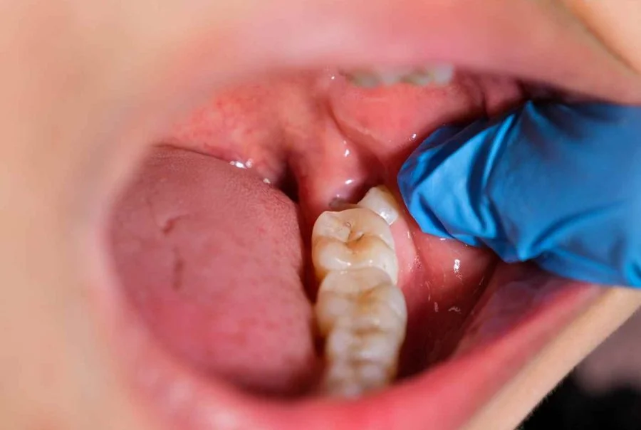
After this information is recorded, a detailed examination of the teeth is performed. Because pathologies that may occur in the teeth are close enough to be reflected in this area. Also, the main result of the chewing system is to bring the teeth together; therefore, a detailed analysis of the overlapping relationship of the teeth, which we call dental occlusion, is extremely important. This is why the treatment of the temporomandibular joint and muscles is managed by specialist dentists.
Imaging Methods in Jaw Pain: Jaw MRI and CT Scan
As a final step, MRI (magnetic resonance imaging) or CT scan is requested when necessary. MRI is the only way to image the temporomandibular joint disc. The disc, which acts like a cushion between the upper and lower jaw and enables jaw movements during function, is directly affected in temporomandibular joint (TMJ) disorders, and MRI imaging is the way to see this disc in detail.
If the solution to the intra-articular pathology is interventional (such as joint surgery, intra-articular lavage/arthrocentesis), MRI imaging may be requested.
The ideal TMJ treatment process involves the joint management of the process by an oral surgeon and a prosthetic dentist. In some cases, physical therapy support may be necessary.